Whether they are contained within the human body or outside of it, 3D printed cells require an enormously complex system of nutrition. Great headway is being made on this front though, and we’re following all of it, with recent strides being made with innovations like new bioprinting technology and the actual creation of blood vessels.
And while so much is happening on the bioprinting front it’s often challenging to keep up, one thing is for sure: this is a fascinating area of discovery. Now, researchers at the Wyss Institute for Biologically Inspired Engineering at Harvard University and the Harvard John A. Paulson School for Engineering and Applied Sciences (SEAS) have developed a method for bioprinting stronger structures in the form of thick vascularized tissue. These constructs are made up of quite the recipe too: human stem cells, an extracellular matrix, and circulatory channels lined with endothelial blood vessel cells.
All of these new processes have been outlined and just published in the Proceedings of the National Academy of Sciences, in the paper ‘Three-dimensional bioprinting of thick vascularized tissues,’ by David B. Kolesky, Kimberly A. Homan, Mark A. Skylar-Scott, and Jennifer A. Lewis.
“This latest work extends the capabilities of our multi-material bioprinting platform to thick human tissues, bringing us one step closer to creating architectures for tissue repair and regeneration,” says Jennifer A. Lewis, Sc.D., senior author on the study.Through vascular ‘plumbing’ which includes both the living cells and extracellular matrix, the scientists are able to create tissue which is sustainable—in fact, they have now been able to offer these structures as viable and functioning for a stunning and unprecedented timeframe of up to six weeks so far. And the secret is, of course, all in the materials.
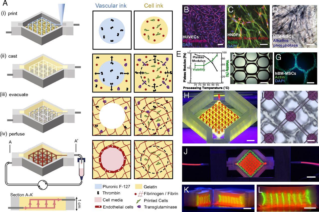
“Central to the fabrication of thick vascularized tissues is the design of biological, fugitive, and elastomeric inks for multimaterial 3D bioprinting,” states the team in their paper.Regarding the ink and the matrix, they go on to state that with their new approach, arbitrarily thick tissues can be bioprinted successfully as the extracellular matrix does not require UV curing and can also be ‘readily expanded’ to other biomaterials too, such as fibrin and hyaluronic acid.
So far, the team has been successful in bioprinting with tissue that is one centimeter thick. They were able to pump bone growth factors through the structures (actually lined with the same endothelial cells found in our blood vessels) allowing for development of cells in a four-week duration.
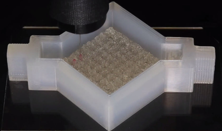
This new bioprinting process is accomplished with a customized silicone mold used as a vehicle for housing and ‘plumbing’ the 3D printed tissue. The researchers were able to print the network of vascular channels, and then next layer live stem cells over that. With each layer, the structure grows and in this process, becomes strong enough to ‘live.’
“This research will help to establish the fundamental scientific understanding required for bioprinting of vascularized living tissues. Research such as this enables broader use of 3D human tissues for drug safety and toxicity screening and, ultimately, for tissue repair and regeneration,” explains Zhijian Pei, National Science Foundation Program Director for the Directorate for Engineering Division of Civil, Mechanical and Manufacturing Innovation, which funded the project.Strength and sustainability in the inks is what makes a substantial difference, allowing for a network of intersections with vascular pillars, microvessels, and post-printing, a cellular liquid which fills in any open regions and links the entire structure together. The result is a structure of soft tissue with the required blood vessels. Both an entry and exit are available on the ‘chip,’ allowing—in an absolutely crucial process–for the proper nutrients necessary to keep the cells alive.
“Jennifer and her team are shifting the paradigm in the field of tissue engineering based on their unique bioprinting approach. Their ability to build living 3D vascularized tissues from the bottom-up provides a potential way to form macroscale functional tissue replacements that can be surgically connected to the body’s own blood vessels to provide immediate perfusion of these artificial tissues, and thus, greatly increase their likelihood of survival. This would overcome many of the problems that held back tissue engineering from clinical success in the past,” said Wyss Institute Founding Director Donald Ingber, MD, PhD.With prefabricated vasculatures, these researchers are able to improve the functioning of the bioprinted cells at their most central point—allowing them to exert influence over what the cells do with substances like growth factors.
Reflecting on this very successful study and research project, the scientists stated that with the ability to make these 3D printed tissues, the ‘exploration of emergent biological phenomena’ is made possible.
“Our 3D tissue manufacturing platform opens new avenues for fabricating and investigating human tissues for both ex vivo and in vivo applications,” concludes the team.Source: http://3dprint.com/123181/3d-printed-vascular-tissues/